









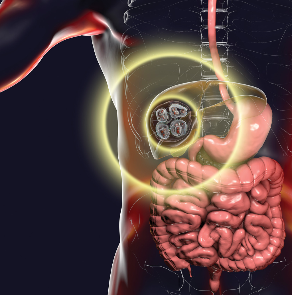
An 82-year-old woman presenting with recurrent severe upper abdominal pain was found to have multiple hydatid cysts involving the liver, lungs, and brain. When follow-up imaging showed enlargement of a liver cyst and concern about possible rupture, laparoscopic decompression offered a minimally invasive approach to managing the largest accessible cysts. Dr Mohammad Arif presents the case and its successful surgical outcome.
Dr Mohammad Arif
MBBS, FRCS (Ireland), FCS (SA)
General and laparoscopic surgeon
Melomed Mitchells Plain Hospital, Cape Town
Case presentation
An 82-year-old woman came with upper abdominal pain in January at Melomed Mitchells Plain, in Cape Town. She was previously seen by a GP in 2024, but we could not access that information. Initial ultrasound followed by a CT scan of her abdomen confirmed multiple hydatid cysts in the lungs, liver, and some degenerative cysts in the brain. Her eyes were cleared by an ophthalmologist. A gastroscopy revealed active gastritis with a small hiatus hernia.
Her abdominal pain settled and she was discharged with Omeprazol for two weeks and Albendazol for six months to treat her gastritis and hydatid disease respectively. She had no chest complaints at that stage.
Her conditions were explained to her, and, given her age, a decision was made to manage her conservatively.
She returned in March with severe upper abdominal pain and was unable to sit upright comfortably. Follow-up ultrasound indicated an increase in the size of one of the cysts in the liver.
After risk assessment and involving the family, a decision was made to operate through laparoscopy and only address the cysts that were very big and easily accessible.
Laparoscopic decompression
A successful laparoscopic decompression of three larger cysts (Figs 2, 3) was done on admission, as there was strong clinical suspicion that the larger cyst, which appeared to be bulging in the epigastric area, might rupture (Fig 1).
This is an operation that is normally done by opening the abdomen in most centres.
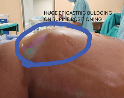
Figure 2: Laparoscopic view of large right lobe liver cyst.
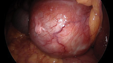
Figure 3: Large left lobe liver cyst, actually an extension from the right lobe cyst.
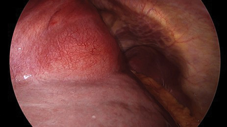
Figure 4: Multiple other liver cysts.
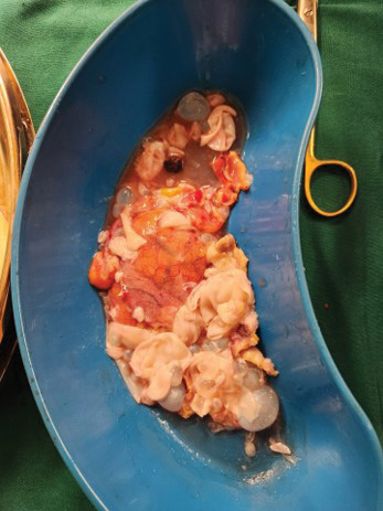
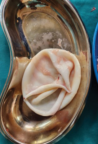
Figure 5: Large hydatid cyst.
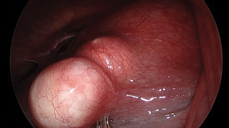
Figure 6: Multiple daughter cysts of different sizes.
The patient did very well, and she will continue Albendazole to complete six months of treatment and will be followed up.
Discussion
Hydatid disease is most common in farming areas. In Cape Town, most patients with the disease come from the Eastern Cape.
Management options include:
- Antiparasitic drugs like Albendazol
- Minimally invasive puncture techniques (PAIR)
- Open or laparoscopic surgery
- Careful ‘watch-and-wait’ approach for inactive cysts.
Experience with emergency decompression is limited, but it is a good and safe option in experienced hands.
South African perspective
Cystic echinococcosis remains an important but poorly characterised zoonotic disease in South Africa, with limited local data on its epidemiology and management.¹,² Recent evidence from the Eastern Cape provides valuable insight into its clinical burden.
A four-year retrospective study at Frere Hospital identified 56 patients treated for liver cystic echinococcosis between 2019 and 2022. Abdominal pain was the most common presenting symptom, reported in 69.6% of patients, while 64.3% presented with a palpable abdominal mass. Disease was confined to the liver in 62.5% of patients, and the right hepatic lobe was affected in 67.9%.¹
Management of hepatic cystic echinococcosis needs to be individualised according to cyst characteristics, anatomical involvement, complications, and the patient’s clinical condition.¹,² In the Eastern Cape cohort, 64.3% of patients underwent surgical partial cystectomy. The investigators reported good short-term outcomes when partial cystectomy was combined with medical therapy, although perioperative bile leak was the most common complication.¹
Contemporary management also includes antiparasitic therapy, percutaneous techniques such as PAIR, surgery, and observation in appropriately selected inactive cysts, reinforcing the importance of tailoring treatment to the individual patient and cyst.²